

Biomedical Engineers in Ultrasound Imaging- perspectives of a Clinical Application Specialist
Ultrasound Imaging
Medical Imaging is one of the most emerging field in this era. Medical Ultrasound is an extensively used medical imaging equipment in the routine clinical workflows for diagnostic purposes. Ultrasound Imaging technology falls into two categories – Diagnostic & Therapeutic Ultrasound
A) Diagnostic ultrasound
“Diagnostic ultrasound is one of the non-invasive imaging techniques that are used to image the anatomy inside the human body. This imaging technique uses ultrasound probes called transducers that produce sound waves with a frequency above 20KHz. Ultrasound probes are either placed on skin or maybe placed inside the body through vagina, GI tract and blood vessels. This is done so as to have a better image quality. The diagnostic can be further classified into Anatomical Ultrasound & Functional Ultrasound.
Anatomical Ultrasound is used to produce images of internal organs or other structures. Whereas Functional Ultrasound uses information such as movement and velocity of tissue or blood, softness and hardness of tissue and other physical characteristics and combines with anatomical images to create information maps.
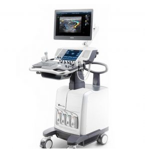
Figure 1:Mindray DC-70 Ultrasound machine
B) Therapeutic Ultrasound
Therapeutic Ultrasound, like diagnostic ultrasound uses sound waves above 20KHz, but does not produce images. It’s purpose is to interact with tissues in the human body. The tissues are either modified or destroyed like heating up of tissue, dissolving blood clots etc. This is made possible with the help of HIFU that can destroy diseased or abnormal tissues such as tumors.”
Working Principle of Ultrasound
“Ultrasound waves are produced by the transducer which can emit ultrasonic waves as well as detect ultrasound echoes that are reflected back.Active elements in the transducers are made of special ceramic crystals called piezoelectrics. These materials have the ability to produce sound waves when an electric field hits them and vice versa. The transducer sends out sound waves into the body. The sound waves are reflected back to the transducer by boundaries between dif erent types of tissue. Echoes on hitting the transducer generates electrical signals that are sent to the ultrasound scanner. The ultrasound scanner calculates the distance from the transducer to the tissue boundary by using the speed of sound and time of each echo’s return. The two dimensional images of tissues and organs are generated using the calculated distances. This forming ultrasound image.
Application of gel on the skin during an ultrasound exam is to reduce air pockets-forming between the transducer and the skin which can block ultrasound waves from passing into the human body.”
Responsibilities of a Biomedical Engineer in Ultrasound Imaging
Biomedical Engineering is the rising field in medical science by involving the knowledge of biology and medicine in combination with the principles of engineering to develop devices and procedures which can solve the greatest number of medical and health related problems in the modern world. Many Biomedical Engineers soon after graduation are taking up the position of Clinical Application Specialist in Ultrasound imaging.The major advantage of a biomedical engineer becoming an application specialist is that they will be able to deliver both clinical as well as technical knowledge to the end users. Hands on scanning experience is an added advantage. Once they are expert in one imaging modality, they can switch into any other modality.
I’m a Clinical Application Specialist of Mindray Ultrasound. I did a Bachelor of Technology in Biomedical Engineering. Soon after my graduation I came to this field through campus placement. So, I didn’t have much idea on how to approach a customer and demonstrate the machine. I was so lucky enough to get trained under some extraordinary persons and I still consider them as the encyclopedias of Ultrasound Imaging. After completing one month of extensive training, I started going to the field and conducting demonstrations. There is a famous saying “Practice makes man Perfect”. I’m fortunate enough to have a mentor, Mr.Maniraj who used to tell us that we should always go to the field and make our hands dirty. His words struck my heart and as a disciple I always implemented that.
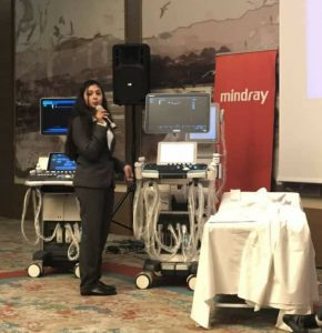
Figure 2: Mindray Turkey Training
At that time I got an opportunity to go to Turkey to have advanced training on Elastography, Radiology, General Imaging, OB/GYN, POC and Cardiology. No words in this entire universe are enough for me to explain how well that training has been conducted and how well it has influenced me. I was fortunate enough to bag second rank when they conducted an examination and while returning back to Dubai, I understood this is the career that I want to pursue further. After that I started learning more and more about Ultrasound, the latest technologies, new inventions etc.
The job of an application specialist is not monotonous. We will get to meet a lot of people on a daily basis and we get to work with them. Everyday when we go to the field we will experience and learn something new from the end users. The major challenge that I faced as an Application Specialist is that since I am dealing with Mindray Ultrasound, the end users mainly in the Middle East Region have a mindset that it is a Chinese brand and the machines are not reliable. It took a while to convince the end users and to change their mindset. The only way to change their mindset is to have continuous demonstrations. We used to conduct workshops for the end users for them to have a hands on experience with the machine. Slowly we changed their mindset and started installing Mindray Ultrasound Machines.
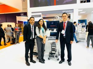
Figure 3:Arab health 2020
Now I can proudly say that we are successful in the UAE market and competing with the major brands like GE, Philips, Siemens, Toshiba, Samsung, Hitachi, Essoate, Sonocite, Sonoscape etc. We should always be pleasant with the end users and should listen to their actual requirements. We need to be with them till the demonstration gets over. It can go upto two weeks to two months. Image optimisation is the main part of the demonstration.
Since ultrasound is user dependent, the acceptable levels of the image quality will differ from doctor to doctor. It’s very important to optimise the images for the end users and moreover we should have a strong clinical background. Through continuous learning only we will be able to achieve this. We should be in a position to understand what images we are seeing on the screen because then only we will be able to optimise it effectively. GE and Philips are the major competitors of Mindray.
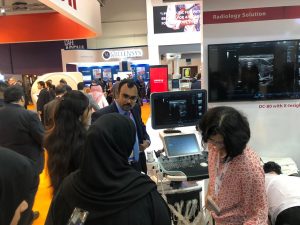
Figure 4:Arab health 2020 Equipment demonstration
Regulatory affairs associated with Ultrasound imaging
The general regulatory affairs associated with Ultrasound imaging are ISO 29821:2018 (Condition monitoring and diagnostics of machine:Ultrasound — General guidelines, procedures and validation), IEC 60601(Electrical safety guidelines), IEC 61000 Electromagnetic compactability (Emission tests: Harmonic & voltage fluctuations and Immunity tests: Conducted & radiation tests). This specifies methods and requirements for carrying out ultrasonic examination of machines, including safety recommendations and sources of error, and provides information relative to data interpretation, assessment criteria and reporting. The regulatory compliances are ensured during the installation phase followed by the Clinical Application Training to the respective department, which emphasizes on the clinical significance of the equipment.
Top tier companies in the Ultrasound imaging field are GE, Philps, Siemens, Samsung, Toshiba, Hitachi and Sonocite. Those who like to travel, I would suggest this is the best field you can choose provided you should have a strong passion towards the Imaging field. Since Ultrasound doesn’t produce any harmful radiations you don’t need to worry about your health too. Scanning experience is an added advantage because you will have to show the scan while doing the demonstration. If you are a biomedical engineer and want to have a career shift from an Application Specialist to a Sonographer, that can also be done provided you have to pursue RDMS. For all those who wanted to become an Application Specialist, just work for it till you make it. Always drive your passion. If you do so, then undoubtedly success is your’s.
Quote reference link : https://www.nibib.nih.gov/science-education/science-topics/ultrasound
Author details:
Aardra Raj
Designation – Clinical Application Specialist of Mindray Ultrasound
2018-Biomedical Engineering graduate
Mail Id- aardraraj97@gmail.com
Contact info- +971567549841


Good read-on for budding aspirants of biomeds interested in ultrasound imaging:)
Congrats aardra!
It was an absolutely subjective and comprehensive presentation that threw light into the intricacies of Bio Medical Engineering exploring new vistas in the arena, which is sure to inspire very many researchers and aspirants in the field. Expecting more from the author again!
Thank You Ma’am for this wonderful blog. This will help many student aspirants like me, who are doubtful about what to do next after completing their BTech degree. I am a one who loves to know more about Biomedical Equipments and exhibit my knowledge in the best possible way. Thank you so much for being my eye opener. I would be much obliged to know more about your journey which will greatly influence my career plans.